Prenatal care, cancer treatment, brain research – these vastly different areas of the healthcare industry have a few things in common.
The first is the technological transformation healthcare has undergone in the past few decades. The second is the fact that all will need an influx of new professionals as the Baby Boom generation retires. The third is they all employ the medical specialty of radiology.
Radiography uses imaging technology to look inside the body. X-rays, MRIs, and ultrasound are some of the most familiar types of radiologic technologies. Radiography provides imaging for radiologists to diagnose and treat patients.
Although radiology is a unique field and requires specialized education, it is integral to a wide variety of procedures across the healthcare industry. Everything from preventive screenings to sports medicine, from assessing a broken bone to conducting regular checkups during pregnancy – all rely on the skill of radiologic technologists.
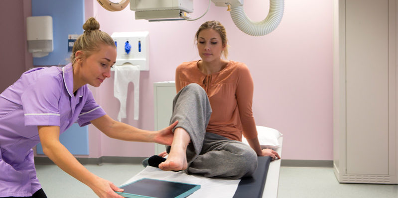
X-rays
X-rays are the most iconic of the medical imaging technologies. Dating back to 1895, they are also the oldest. In fact, German physics professor Wilhelm Conrad Röntgen coined the ionizing radiation an “X ray” because it was unknown at the time. Just a year after this discovery, medical testing had incorporated the technology.
It is the radiation of the X-ray that gives radiologists and radiographers of today their job titles – though the profession and the procedures themselves are infinitely safer than in the early days before radiation was well understood.
Radiography now incorporates other imaging techniques using sound and magnets, but X-rays are still the predominant imaging test (and the second most common medical test after blood tests) in healthcare. Mammograms, dental care, and arthritis treatment are just a few of the procedures relying on X-ray technology.
MRI scans
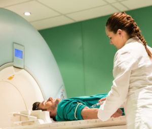
A magnetic resonance imaging (MRI) scanner works by forming a strong magnetic field around the body of a patient. The magnet’s energy is picked up by hydrogen atoms in the body’s water molecules. The “excitement” of these atoms creates a radio signal which is recorded and measured by the equipment and interpreted as an image or a series of images. Differences in tissues are shown in color or contrast differences, giving the medical team a precise picture of what is going on internally.
When the MRI scanner was invented in 1971, it kickstarted a revolution in cancer detection and new ways of understanding brain functions. By the end of the decade, the technique had been refined from taking a few hours to a few seconds. Also, testing had moved from mice to humans. In 1980, doctors used an MRI to detect tumors, cancer, and liver problems in a patient for the very first time.
Today, MRIs are sophisticated enough to identify thin spots in artery walls, metabolism imbalances, and even the delicate activity of neurons in the brain.
Sonography
Is it a girl or a boy? Sonography is usually associated with the ultrasounds produced during pregnancy – those black and white photos of a developing baby. But sonography – a non-invasive imaging procedure using high-frequency soundwaves – is used to examine virtually the entire body to look for abnormalities. This includes the brain, circulatory system, and other soft tissues.
Technological advances have made the images less grainy and more precise, and cut down on the need for more invasive procedures. Unlike MRIs or X-rays, sonograms are unique in that they allow the patient to move around and can produce images in real time to see the body in motion.
Bright outlook
Radiography is one of the fastest growing careers in health care, driven by an aging population and the development of advanced treatments for cancer and other conditions. Some specialized positions require graduate or medical degrees.
Established in 1997, the HFC Radiographer (RAD) program spans 22 months with students learning in classroom and clinical settings concurrently. HFC RAD graduates are instructors both in the classroom and at the clinical sites.
“The beauty of our field is the opportunity for graduates to cross-train in other imaging modalities, such as CT, MRI and mammography. There are many different career paths our graduates can take,” said Sharon Wu, HFC RAD program director.
According to Wu, employers in southeast Michigan value and hire graduates of the HFC RAD program. The odds are good that if you have an imaging procedure done in the Metro Detroit area, an HFC RAD graduate will be performing it.
Graduates from the HFC Radiographer program have a 93 percent first-time pass rate on the American Registration of Radiologic Technologists (ARRT) National Credentialing Exam over the last five years. Of the students who took the national exam, 103 out of 111 passed on the first try from 2012-16.
For questions about the HFC Radiographer Program, contact Wu at 313-317-6595 or [email protected].
Visit hfcc.edu/academics/programs/radiographer.
Naomi Sheehan & Kurt Anthony Krug